Congenital Heart Surgery (Common Operations — Palliative, Single-Ventricle Staged & Biventricular Repairs)
Source: Norwood WI et al. NEJM 1983; Single Ventricle Reconstruction (SVR) Trial (Ohye RG et al., NEJM 2010); Jatene AD et al. JTCVS 1976; Fontan & Baudet Thorax 1971. โครงเรียงตาม operation (palliative → single-ventricle staged → biventricular corrective) — แต่ละ operation ระบุว่าทำอะไร + ใช้กับโรคไหน
🟡 กลุ่ม A — Palliative / Temporizing Procedures
operation กลุ่มนี้ไม่ได้แก้โรคให้หาย แต่ปรับ pulmonary blood flow ให้สมดุลเพื่อประคองจนโตพอทำ definitive repair
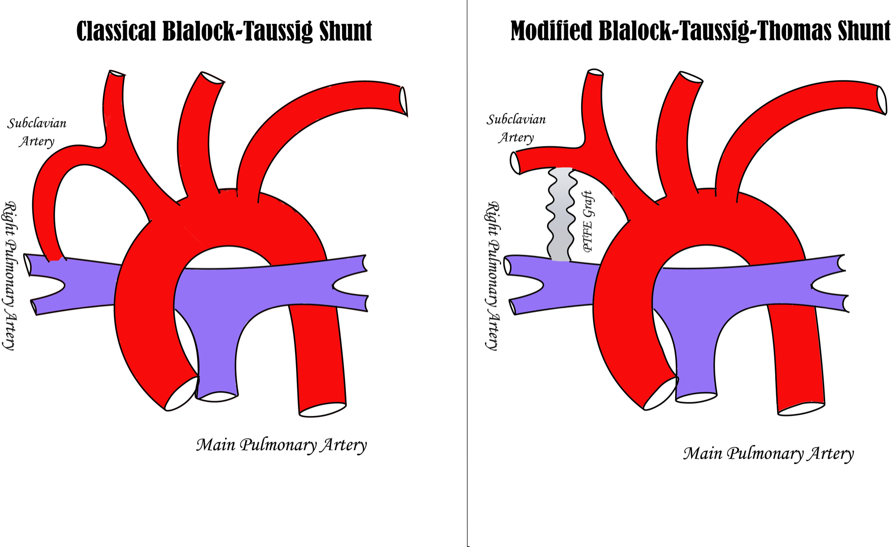
1. Modified Blalock–Thomas–Taussig (BTT) shunt
ทำอะไร: สร้าง Gore-Tex (PTFE) conduit เชื่อม subclavian หรือ innominate artery เข้ากับ pulmonary artery ส่งผลให้เกิด controlled left-to-right shunt ที่เพิ่ม pulmonary blood flow
ใช้เมื่อ: pulmonary blood flow น้อยเกินจน cyanosis รุนแรง — tetralogy of Fallot ที่มี severe RVOT obstruction, pulmonary atresia, หรือ tricuspid atresia ระยะแรกก่อนเข้า single-ventricle pathway เนื่องจากการเพิ่ม flow ทำให้ oxygenation ดีขึ้นและเปิดโอกาสให้ pulmonary arteries โตพอสำหรับ repair ภายหลัง
2. Pulmonary Artery Banding (PAB)
ทำอะไร: รัด main pulmonary artery เพื่อจำกัด pulmonary blood flow (ทิศตรงข้าม BTT shunt)
ใช้เมื่อ: excessive left-to-right shunt ทำให้ pulmonary overcirculation — large VSD หลายรู, single ventricle without pulmonary stenosis, AV canal defect ที่ยังไม่พร้อม repair — การปล่อยให้ flow มากเกินนานจะเกิด pulmonary vascular disease (Eisenmenger) ที่ irreversible ดังนั้น band จึงป้องกัน pulmonary bed ไว้
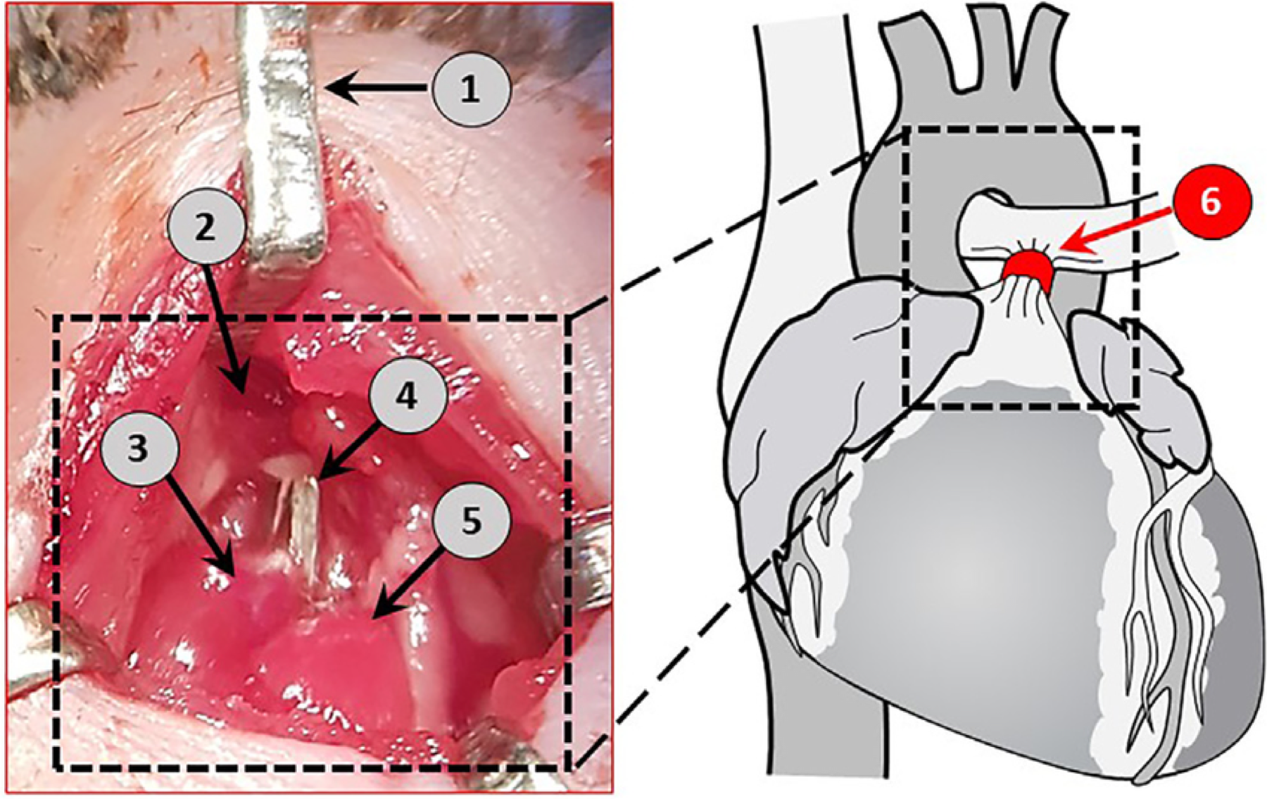
Intraoperative illustration of the pulmonary artery banding 1) chest retractors, 2) aortic arch, 3) pulmonary trunk, 4) ligating clip placed around the pulmonary trunk, 5) left atrium, 6) a schematic representation of the pulmonary trunk constriction.
3. Adjuncts ก่อนผ่าตัด
-
Prostaglandin E1 (alprostadil) infusion — คง ductus arteriosus ให้เปิดใน ductus-dependent lesions เนื่องจาก PGE1 คลาย ductal smooth muscle ทำให้ systemic/pulmonary flow ยังพึ่ง ductus ได้จนกว่าจะผ่าตัด
-
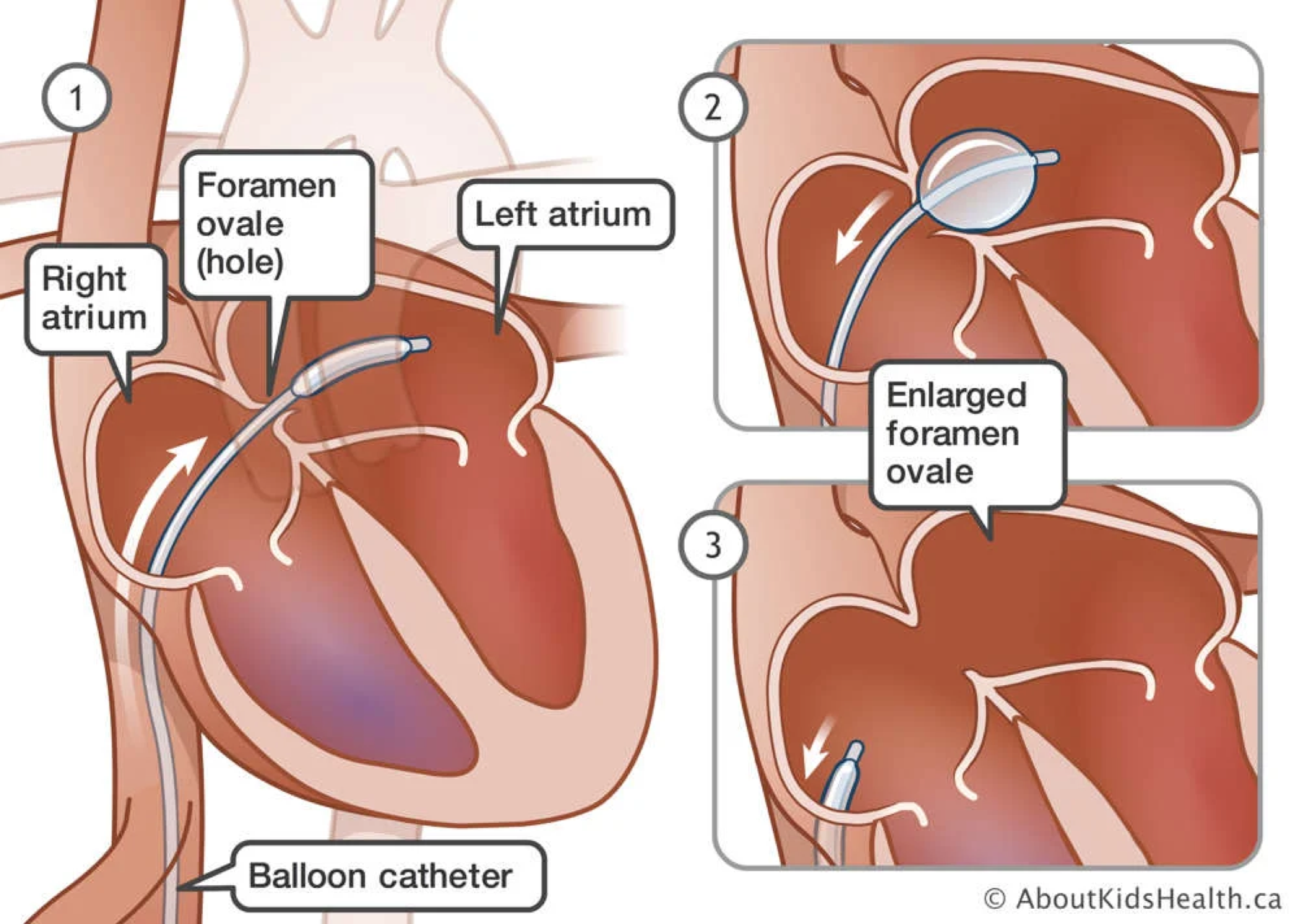
Balloon atrial septostomy (Rashkind) — เพิ่ม intracardiac mixing โดยเฉพาะใน transposition of great arteries (TGA) ที่ต้องอาศัย mixing ประคอง oxygenation ก่อน arterial switch
🔵 กลุ่ม B — Single-Ventricle Staged Palliation
ใช้เมื่อมี functional ventricle เพียงห้องเดียว (HLHS, tricuspid atresia, double-inlet ventricle) — สร้าง two-pump circulation ไม่ได้ จึงให้ single ventricle สูบไป systemic ส่วนเลือดไป lung อาศัย passive venous flow — ทำเป็นสามขั้นเพื่อให้ pulmonary vasculature ปรับตัว และพึ่ง low PVR อย่างยิ่ง
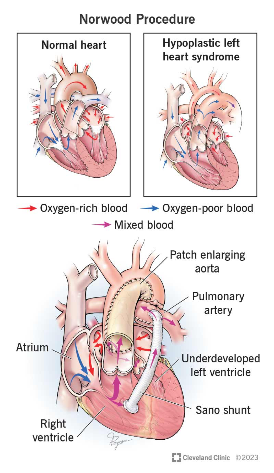
4. Stage I — Norwood Procedure
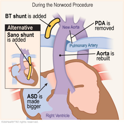
ทำอะไร: ทำในทารกแรกเกิด — สร้าง neo-aorta จาก single ventricle (รวม main PA กับ hypoplastic aorta), atrial septectomy ให้ mixing, และสร้าง shunt ไป PA — เลือกได้ modified BTT shunt หรือ Sano shunt (RV-to-PA conduit)
ใช้เมื่อ: hypoplastic left heart syndrome (HLHS) เป็นข้อบ่งชี้คลาสสิก + single-ventricle variants ที่มี systemic outflow obstruction
5. Stage II — Bidirectional Glenn (BDG) / Hemi-Fontan
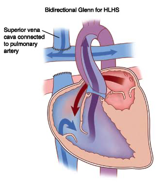
Bidirectional Cavopulmonary shunt
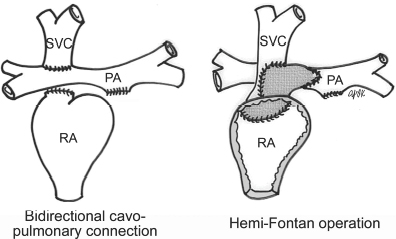
ทำอะไร: ต่อ SVC เข้า pulmonary artery โดยตรง และ occlude shunt เดิม — เลือด deoxygenated จากร่างกายส่วนบนไหลเข้า lung แบบ passive ไม่ผ่าน ventricle → ลด volume load ของ single ventricle (Hemifontan คือ Glenn ที่เอา patch มาปิด RA ไว้ ไม่ต้องเย็บปิด พอขยับไปทำ fontan เต็มก็แค่ต่อรูเดิม แต่ตอนนี้ไม่ทำละ)
ใช้เมื่อ: ทำ 3–6 เดือนหลัง Norwood (SVR Trial → mortality ต่ำสุดช่วงนี้) — ต้องมี low PVR
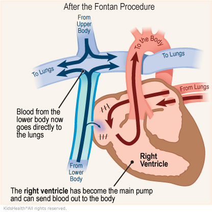
6. Stage III — Fontan (Total Cavopulmonary Connection)
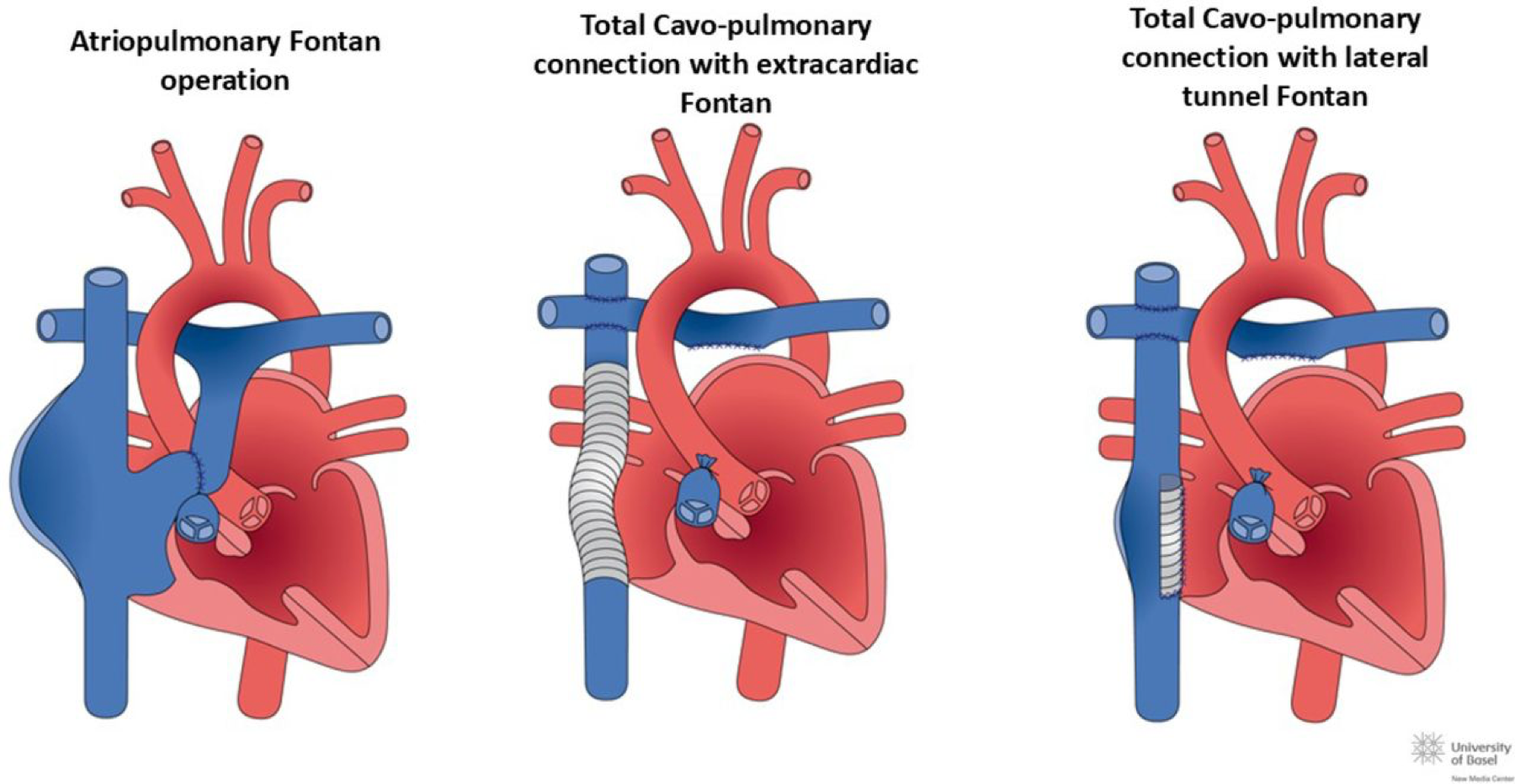
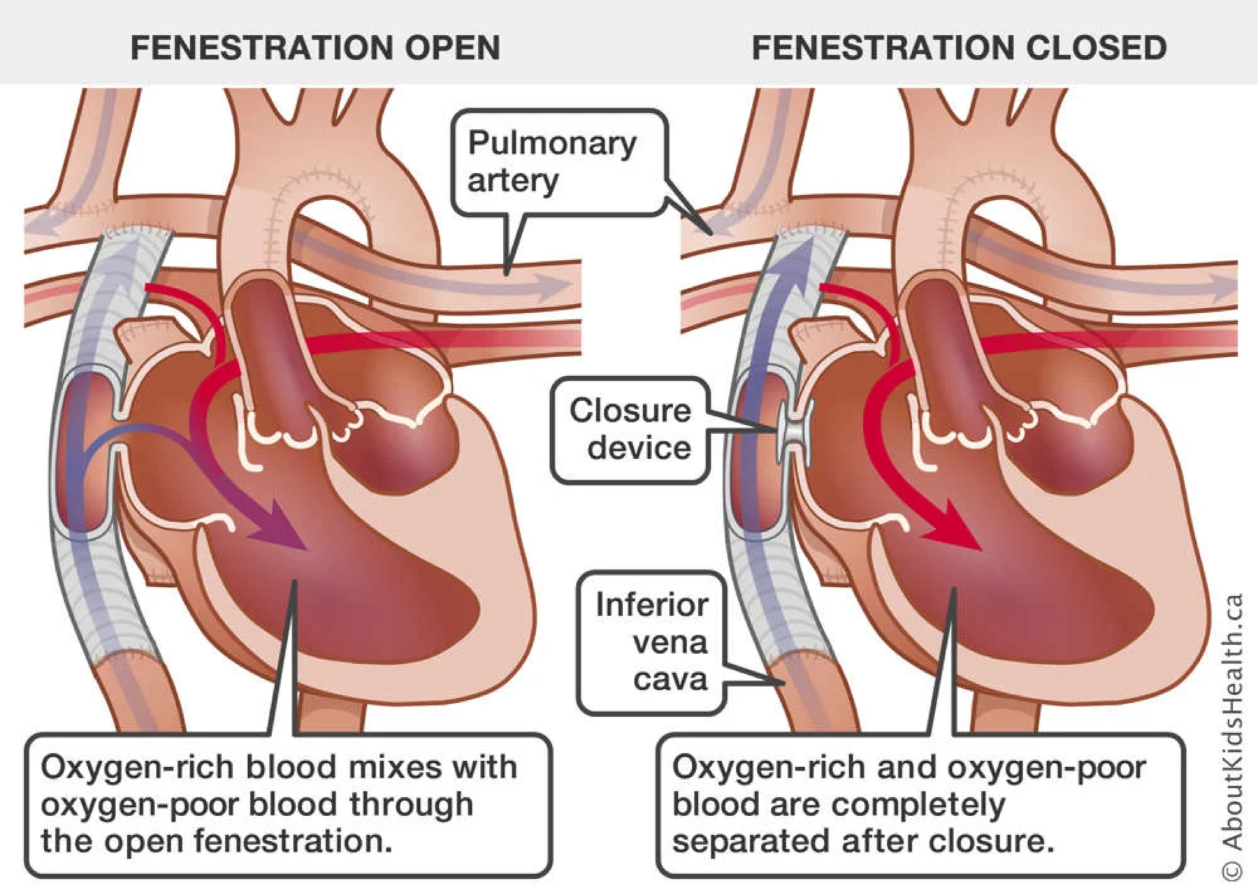
ทำอะไร: ต่อ IVC เข้า pulmonary artery — completion ของ total cavopulmonary connection (extracardiac conduit หรือ lateral tunnel) → systemic venous return ทั้งหมดไป lung โดยไม่ผ่าน ventricle → series circulation สมบูรณ์; บางรายทำ fenestration เป็น pop-off ลด Fontan pressure
ใช้เมื่อ: completion ของ single-ventricle pathway อายุ 2–4 ปี — ภาวะแทรกซ้อนระยะยาว: protein-losing enteropathy, plastic bronchitis, Fontan-associated liver disease, atrial arrhythmia (ระบบเปราะบางต่อการเพิ่ม PVR)
Failing Fontan — 2 modes (fellowship point): ระบบ Fontan เสื่อมลงตามเวลาเสมอ แบ่งการ fail เป็น 2 แบบที่พยากรณ์ต่างกัน — (1) impaired ventricular function (IVF) onset ช้ากว่า (median ~8 ปี post-TCPC) มี progressive systolic dysfunction + AV valve regurgitation; (2) preserved ventricular function (PVF) แต่ Fontan circulation fail onset เร็วกว่า (median ~2.6 ปี) มาด้วย PLE / plastic bronchitis — กลุ่ม PVF นี้ ventricular function ยังดีแต่ เสี่ยงตายสูงกว่า IVF >3 เท่าที่ 1 ปี จึงต้อง refer transplant เร็ว. สาเหตุ circulation fail: anatomic obstruction, thromboembolism, elevated PVR, pulmonary AVM, lymphatic dysfunction. ปลายทางคือ heart transplant (post-Fontan transplant survival ~78% ที่ 5 ปี); lymphatic complication (plastic bronchitis/PLE) มี fenestration หรือ innominate-vein turn-down decompress thoracic duct ช่วยได้
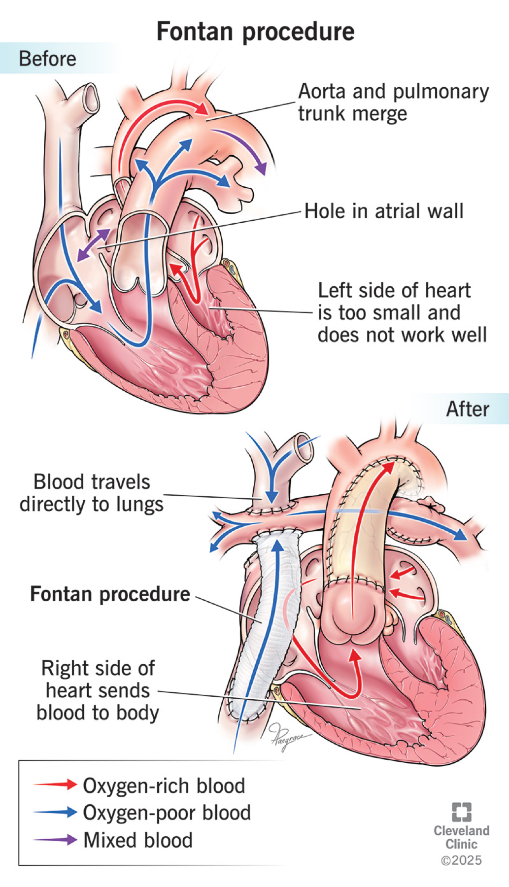
fenestration fontan — ลดปหเวลา PVR สูง ๆ จะได้มี flow เข้า heart
🟢 กลุ่ม C — Biventricular Corrective Repairs
ใช้เมื่อมี ventricle สองห้องใช้งานได้ทั้งคู่ — คืน circulation เป็น series circuit ปกติ โดยปิด shunt ผิดปกติและแก้ obstruction
7. ASD Closure
ทำอะไร: ปิด atrial septal defect ด้วย patch (open) หรือ percutaneous device (Amplatzer) → กำจัด left-to-right shunt ระดับ atrium
ใช้เมื่อ: secundum ASD ที่มี significant shunt (Qp:Qs สูง) หรือ RV volume overload; primum/sinus venosus ASD มักต้อง surgical repair
8. VSD Closure
ทำอะไร: ปิด ventricular septal defect ด้วย patch ส่วนใหญ่เข้าทาง tricuspid valve (ไม่ต้อง ventriculotomy) → กำจัด left-to-right shunt ระดับ ventricle
ใช้เมื่อ: large VSD ที่ทำให้ heart failure, failure to thrive, pulmonary overcirculation หรือ recurrent endocarditis; VSD เล็กหลายรายปิดเองได้
9. Tetralogy of Fallot (TOF) Repair
ทำอะไร: แก้สอง defect พร้อมกัน — ปิด VSD ด้วย patch และ relieve RVOT obstruction
-
Transannular patch (TAP): ขยาย RVOT ได้ดีแต่แลกกับ pulmonary insufficiency ระยะยาว
-
Valve-sparing repair (VSR): เก็บ pulmonary valve ผ่าน muscle bundle resection + commissurotomy → ลด pulmonary regurgitation
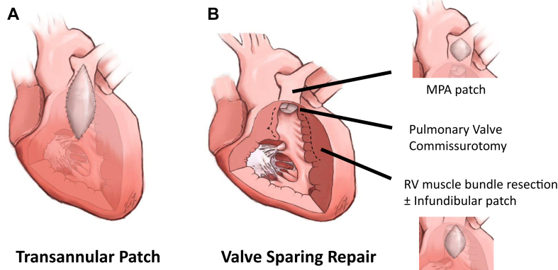
ใช้เมื่อ: tetralogy of Fallot ทุกราย (อายุ 6 เดือน–2 ปี); ผู้ที่ได้ TAP มักต้อง pulmonary valve replacement ในวัยผู้ใหญ่จาก chronic PR
10. Arterial Switch Operation (Jatene)
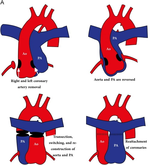
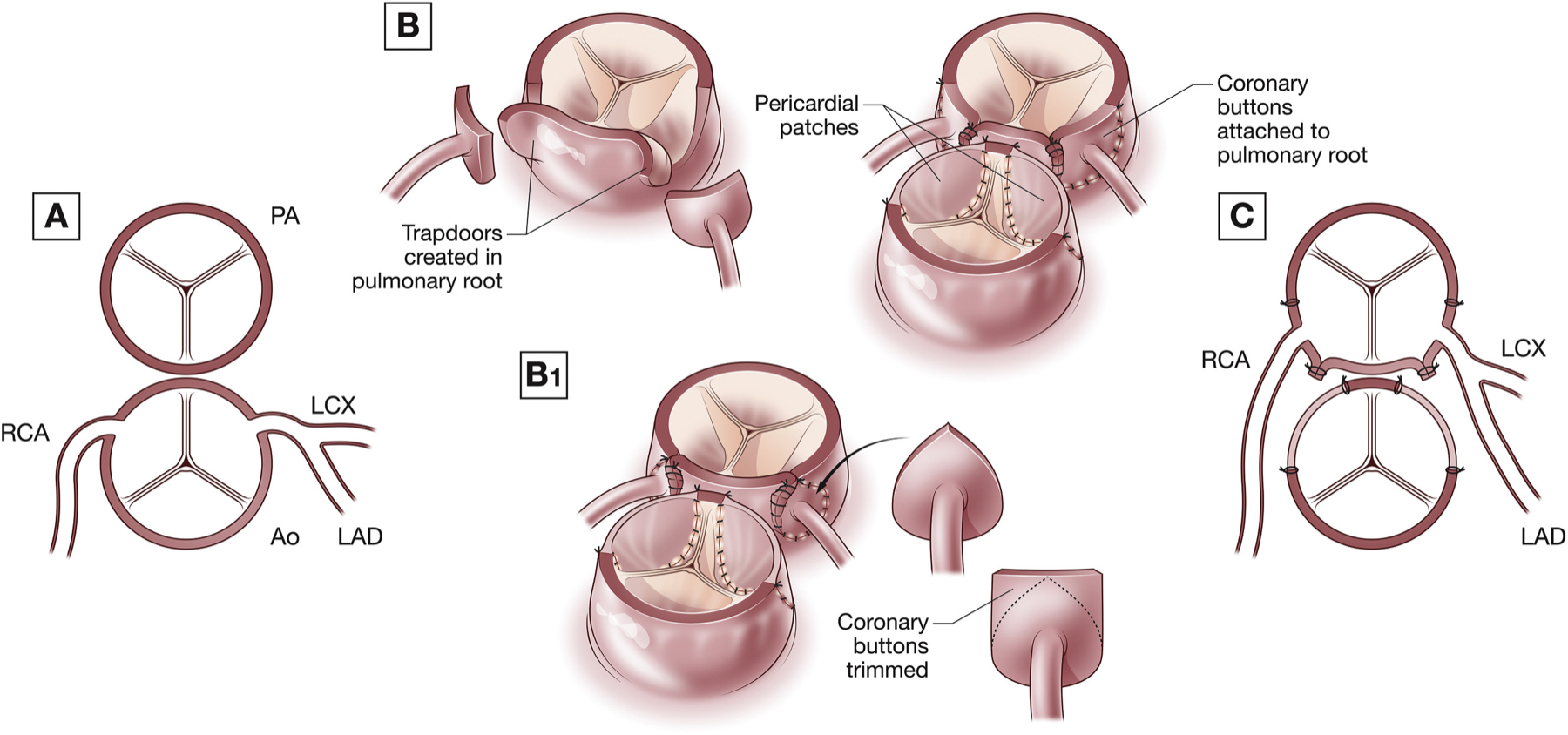
ทำอะไร: ตัด great arteries เหนือ valve แล้วสลับตำแหน่ง aorta กับ PA พร้อม reimplant coronary arteries → anatomic correction ให้ LV กลับมาเป็น systemic pump
ใช้เมื่อ: transposition of great arteries (TGA) ทั้ง intact septum และมี VSD; เป็น operation of choice แทน atrial switch (Mustard/Senning) เดิมที่ปล่อย RV เป็น systemic pump → RV failure
11. Rastelli Procedure
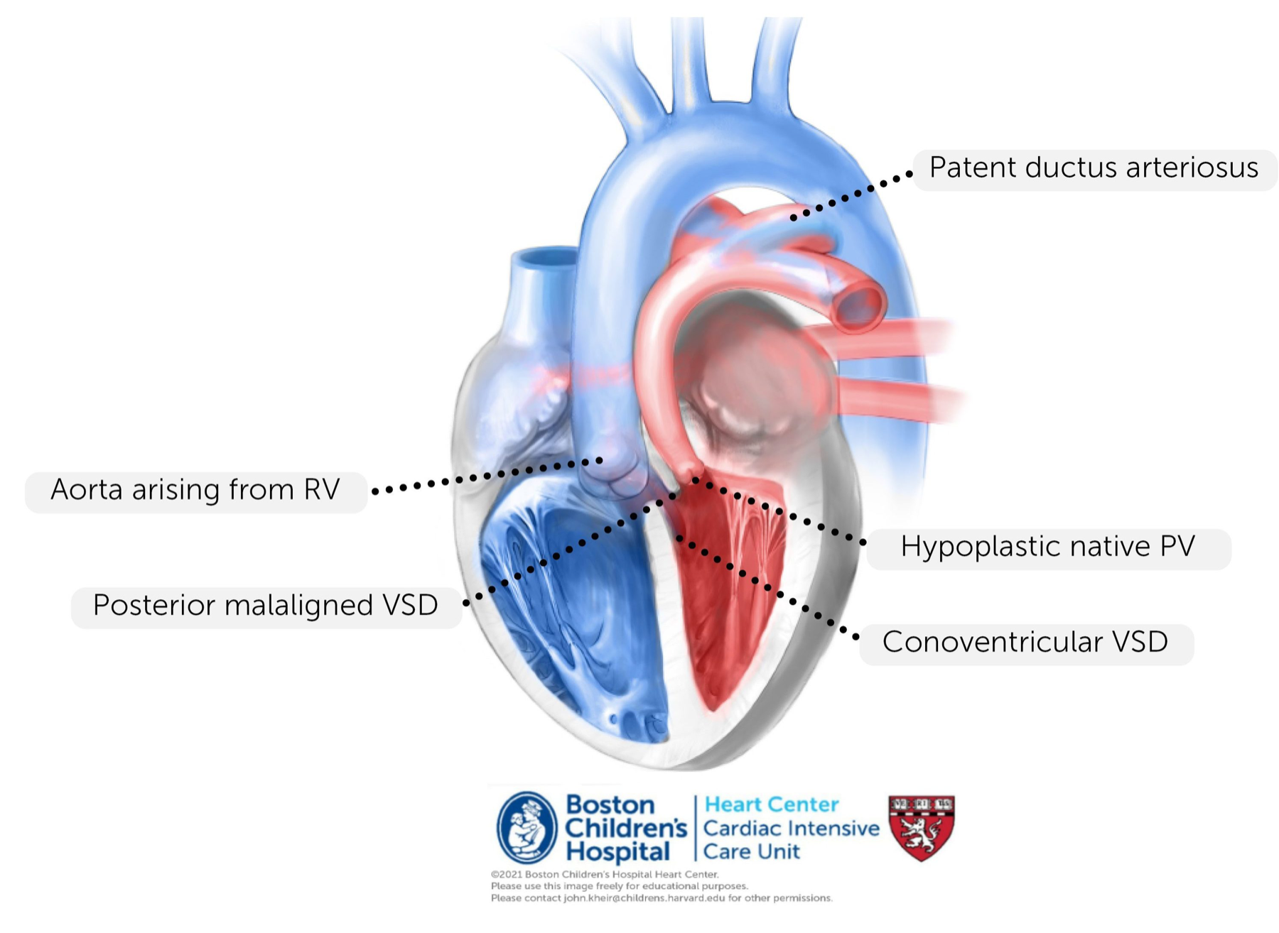
ทำอะไร: baffle เลือด LV ผ่าน VSD → aorta และสร้าง RV-to-PA valved conduit เลี่ยง pulmonary obstruction
ใช้เมื่อ: TGA + VSD + pulmonary stenosis (LVOT obstruction) ที่ทำ arterial switch ตรงๆ ไม่ได้
12. Coarctation of Aorta Repair
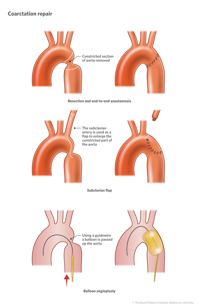
ทำอะไร: ตัด coarcted segment → end-to-end anastomosis หรือ subclavian flap aortoplasty หรือ balloon/stent → ปลด obstruction ของ aortic arch
ใช้เมื่อ: coarctation ที่มี significant gradient หรือ upper-lower limb BP differential; มักทำได้โดยไม่ต้อง CPB (extracardiac)
13. PDA Ligation
ทำอะไร: ผูก/ตัด patent ductus arteriosus (surgery) หรือ device/coil (percutaneous) → กำจัด left-to-right shunt ผ่าน ductus
ใช้เมื่อ: PDA ที่มี significant shunt → pulmonary overcirculation/heart failure โดยเฉพาะ preterm (ลองยา NSAID/indomethacin ก่อน); extracardiac ไม่ต้อง bypass
🩻 การประเมินก่อนเลือก pathway
ใน TOF การประเมินว่า pulmonary arteries โตพอ repair หรือยังใช้ McGoon ratio (branch PA เทียบ descending aorta) และ Nakata index (PA cross-sectional area index ต่อ BSA); single-ventricle pathway การเดินหน้า Glenn/Fontan ต้องมี low PVR เสมอ (physiology พึ่ง passive flow) — เกณฑ์ candidacy ที่นิยมอ้าง: PVRI (indexed) ในอุดมคติ < 2 WU·m², ยอมรับได้ถึง < 3 WU·m², และ PVRI > 4 WU·m² ถือเป็น high-risk/near-contraindication (คลาสสิกจาก Choussat/Fontan “Ten Commandments”); mean PA pressure ที่เหมาะ ~< 15 mmHg
⚠️ ระวังหน่วย (unit trap): ตัวเลขเหล่านี้เป็น PVRI = indexed (WU·m²) — ต่างจาก PVR ธรรมดา (WU) ที่ไม่คูณ BSA. Source extraction (SNC2) ระบุ ”< 4 WU” ซึ่งกำกวมเรื่องหน่วยและเป็นค่า upper-limit/contraindication มากกว่าเป้า candidacy — ค่าที่ควรใช้เป็นเป้าคือ PVRI < 2–3 WU·m². 🔴 ตัวเลขจำเพาะ (2 vs 3 vs 4) แปรตาม institution/era — ยึด congenital team protocol
Landmark references: Norwood WI et al. (NEJM 1983) — staged palliation ของ HLHS; Single Ventricle Reconstruction Trial (Ohye RG et al., NEJM 2010) — BTT vs Sano shunt ใน Norwood; Jatene AD et al. (JTCVS 1976) — arterial switch; Fontan F & Baudet E (Thorax 1971) — Fontan; TOF repair ครั้งแรกสำเร็จ 1954 ต่อยอดจาก Blalock-Thomas-Taussig shunt (1944)
⚠️ เพื่อการศึกษาและ reasoning — ไม่ใช่ surgical decision-making จริง การเลือก procedure/timing/candidacy อยู่ในการดูแลของ congenital cardiac surgery team และ pediatric cardiology เสมอ