IMG_0675
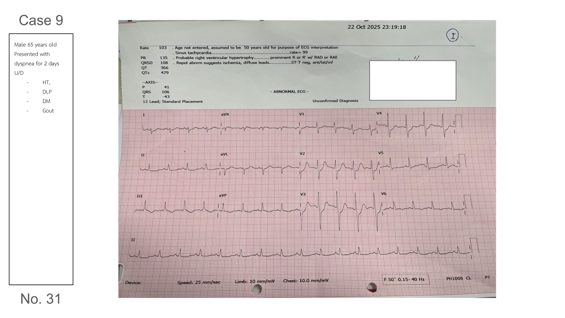
Dx: Posterior wall STEMI
-
describe
sinus tachycardia rate 100
RAD
STD in V2-3 with prominent R wave in V2 — posterior wall STEMI
STE dose not seen in inferior lead, but we have seen inverse T, with the reciprocal STD in lead I, aVL
imp — isolate? posterior wall STEMI, RCA occlusion
**
this need to be differentiate from De winter pattern — by upsloping STD and tall T wave